3D Scanning and printing at the Smithsonian:
Sunday, September 29, 2013
DIGM 620 - Week 2
 |
| Generated using photogrammetry |
Recent years have seen an increase in the adoption of digital tools by paleontologists. What was once a discipline that relied solely on shovels, picks, and brushes has become one that actively utilizes 3D scanning, 3D printing, biomechanical modeling, and animation. Creating 3D models of fossils has opened up many new possibilities for both research and outreach.
On a practical level, digitization can reduce some of the inconveniences and risks of working with fossilized objects. It can minimize the number of times a potentially fragile specimen needs to be handled. It is also much easier to manipulate and pose a large specimen in digital space.
Digital models can facilitate the sharing of specimens between researchers. Previously if a researcher wanted to study a specimen from a collection in another part of the world, he or she would either have to travel to that institution or request that the samples be shipped. Shipping a physical fossil always comes with risks. The advancement of the internet now allows digital models to be easily viewed, hosted, and shared between individuals.
My current focus is on how fossil collections and the exciting science surrounding them can be made more accessible to the public. The fossils on display in a museum are only a small fraction of their total collection. The vast majority of the fossils are sitting in specimen cabinets behind the scenes. Some of these collections are significant findings with wide-reaching implications for the subject of evolution, ecology, development, and more. Due to fragility or impracticality, a great deal of these collections are generally off-limits to the public.
There is a growing movement to digitize collections for preservation, integration of data, and to benefit the public. Many museums and institutions are starting to host their specimen catalogues for public viewing. Some are even sharing 3D models.
C-T scanning and laser scanning are currently the most prominent techniques for bringing a fossil into 3D digital space, but these often require expensive equipment and software. I would like to explore the possibilities of an alternate technique known as photogrammetry. Automated digital photogrammetry software sifts through a series of photographs of an object, picks out overlapping regions, and generates a 3D point cloud which then can be transformed into 3D geometry. Of all the techniques, photogrammetry is currently the most cost-effective way to scan fossils. It is a useful technique because the only major piece of equipment required is a camera, there are several free services available, its resolution is comparable to other techniques, and it generates texture maps in addition to geometry.
I currently have permission to scan a collection of Devonian placoderm fish, called Bothriolepis, at the Academy of the Natural Sciences. This collection will likely play an important role in my eventual thesis project (more on that in a later entry). Having polished digital models of these fossils will be a useful asset down the road.

I will be using Autodesk's free 123D Catch service and software to generate models. I have performed a few guerrilla photoshoots to test out the feasibility of the technique. The two examples are available below (using the service p3d.in)
Here's an eagle statue near the steps of the art museum:
http://p3d.in/D2GFW
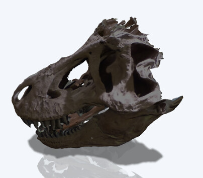
Here's a Chasmosaurus skull from the Academy:
http://p3d.in/VALel
I was surprised by how much it was able to catch. The areas that were in shadow lost a lot of detail though, but could be fixed with proper lighting (The Academy is pretty dim). Also, occluded regions (back of the frill) were lost.
The project will serve to both generate stand-alone digital models, produce assets to serve as reference material for my thesis project, and to gain further familiarity with the photogrammetry work flow.
Some of the challenges I will be facing include: camera-related distortion, capturing fine-details on surfaces, post-work, capturing occluded regions, cleaning up digital artifacts and holes, retopologizing, avoiding 'digital erosion', transferring texture information onto UV-maps, and poly-reduction.
Some of the challenges I will be facing include: camera-related distortion, capturing fine-details on surfaces, post-work, capturing occluded regions, cleaning up digital artifacts and holes, retopologizing, avoiding 'digital erosion', transferring texture information onto UV-maps, and poly-reduction.
- A series (3-4) of digitized 3D fossils presented either through an animated turntable or embedded in a website.
- (tentative) A tutorial video discussing the workflow.
This Week's Activities:
- Meeting with Dave Mauriello to discuss project and research question (1 hr)
- Web stream of iDigBio Paleontology Digitization Workshop (2 hrs)
- Setting up Blog (2 hr)
- Introduction Entry (1 hr)
- Pitch Entry (1 hr)
- David Wagner talk @ ANS regarding American Wildlife Art (1 hr)
- Misc Reading (3 hr)
- First fieldwork day at Inversand dig site (ENVS 865 - Field Methods in Paleontology)
Readings:
Min Zhu et al. "A Silurian placoderm with osteichthyan-like marginal jaw bones". Nature, 2013.
Werdelin, Lars, and John A. Long. "Allometry in the placoderm Bothriolepis canadensis and its significance to antiarch evolution." Lethaia, 1986
Cloutier, R. "The fossil record of fish ontogenies: insights into developmental patterns and processes." Seminars in cell & developmental biology. Academic Press, 2010.
Wells, N. A., and J. A. Dorr Jr. "Form and function of the fish Bothriolepis (Devonian: Placodermi, Antiarchi): The first terrestrial animal." Michigan Academician, 1985.
Janvier, Philippe, Sylvain Desbiens, and Jason A. Willett. "New evidence for the controversial “lungs” of the Late Devonian antiarch Bothriolepis canadensis (Whiteaves, 1880)(Placodermi: Antiarcha)." Journal of Vertebrate Paleontology, 2007
Goujet, Daniel. "“Lungs” in Placoderms, a persistent palaeobiological myth related to environmental preconceived interpretations." Comptes Rendus Palevol, 2011.
All Your Yesterdays: Extraordinary Visions of Extinct Life from a New Generation of Paleoartists, Irregular Books, 2013;
Expected activities:
Coordinate with Dr. Daeschler and plan first photoshoot @ the Academy
Contact Jason - to see if I can get 123D Catch installed on one of the lab PCs.
Young, Gavin C. "Placoderms (armored fish): dominant vertebrates of the Devonian Period." Annual Review of Earth and Planetary, 2010
Friday, September 27, 2013
Introduction

How about an introduction?
Hello, I am Daniel Newman. I am entering the second year of my Digital Media M.S. program. My academic background can best be explained as an odd oscillation between biology and art. For my undergraduate at Providence College I earned a B.S. in Biology with a minor in Studio Art. After college, I worked as a laboratory technician by day but also managed to complete a certificate program in Natural Science Illustration at RISD by night. Unable to choose a career path like a normal person, I seem to have woven two of them together. Here at Drexel University, I hope to leverage my varied experiences and produce some really neat work.
If you would like to see my current creative portfolio, follow this link:
http://www.danieljoelnewman.com
While everyone was applying sunblock and grilling hamburgers this summer, I was spending most of my time searching through tomes of esoteric knowledge in search of a suitable digital media thesis project. I went through several hilariously bad iterations.
At end I found myself returning to a previous interest of interpretive visualization, especially regard to the biological sciences. There are many processes in science that exist beyond our ability to physically see them. Molecules are smaller than the wavelength of visible light and so are entirely invisible to us. Galaxies exist beyond the physical and temporal scales that we typically find comfortable. There were once entirely alien ecosystems full of organisms on this planet that no longer exist. By using digital tools, in my case digital animation, interpretive visualization attempts to make these processes accessible. One can't make them truly visible, but one can hopefully convey important theories, concepts, relationships, and truths that would otherwise be obscure.
The example that I continue to lean on for inspiration is XVIVO and BioVisions' 2005 animation Inner Life of the Cell. Using digital animation, the complexity and dynamism of cellular machinery is explored in what was at the time a uniquely cinematic way. Important biological truths about scale and molecular interaction were made clear to the viewer.
http://www.youtube.com/watch?v=wJyUtbn0O5Y
Subscribe to:
Posts (Atom)